Soft tissue implant dentistry
Soft Tissue–Implant Interface
Early in the modern history of dental implants, most research and clinical focus was devoted to the bone–implant
interface (achieving osseointegration). Little attention was given to gingival health and the architecture surrounding implant components. This has changed such that the peri-implant soft tissues are given strong consideration
during treatment planning and the placement of dental implants. The desire to optimize esthetics after implant
placement is now a key goal of those participating in implant placement and restoration, particularly, for implants
in the maxilla whose gingival margins will be visible while smiling.
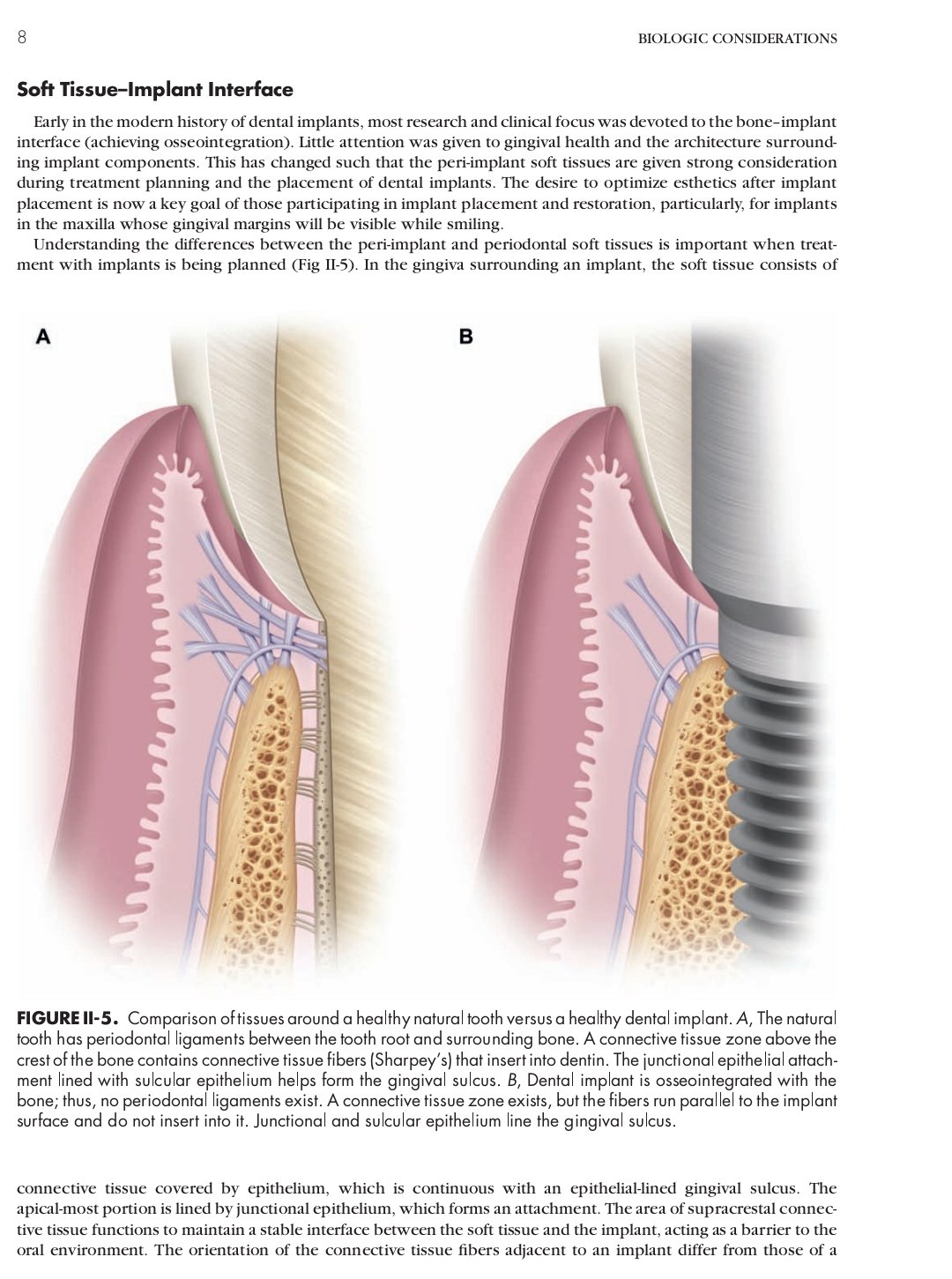
Understanding the differences between the peri-implant and periodontal soft tissues is important when treatment with implants is being planned (Fig II-5). In the gingiva surrounding an implant, the soft tissue consists of
FIGURE II-5. Comparison of tissues around a healthy natural tooth versus a healthy dental implant. A, The natural
tooth has periodontal ligaments between the tooth root and surrounding bone. A connective tissue zone above the
crest of the bone contains connective tissue fibers (Sharpey’s) that insert into dentin. The junctional epithelial attachment lined with sulcular epithelium helps form the gingival sulcus. B, Dental implant is osseointegrated with the
bone; thus, no periodontal ligaments exist. A connective tissue zone exists, but the fibers run parallel to the implant
surface and do not insert into it. Junctional and sulcular epithelium line the gingival sulcus.
connective tissue covered by epithelium, which is continuous with an epithelial-lined gingival sulcus. The
apical-most portion is lined by junctional epithelium, which forms an attachment. The area of supracrestal connective tissue functions to maintain a stable interface between the soft tissue and the implant, acting as a barrier to the
oral environment. The orientation of the connective tissue fibers adjacent to an implant differ from those of a
natural tooth. This area of connective tissue is 1 to 2 mm in height. This becomes important when determining the
health of the peri-implant soft tissues. Probing depths in a healthy implant will typically be 1 to 2 mm less than the
total measured dimension from the crest of the sulcus to the alveolar bone crest.
Another obvious difference between teeth and implants is that teeth have a periodontal ligament with connective tissue fibers that suspends the tooth in the alveolar bone. A well-integrated implant, however, is in direct contact with the bone, without any intervening soft tissue. This difference has a major impact on the biomechanics,
proprioception, and prosthetic considerations for implants versus natural teeth.
You may also like
Proposal for Hotels -2025
10 January, 2025

measurements specific to implant placement
22 December, 2024

Implant Planning Imaging
22 December, 2024